Diagnosing Brain Tumors from MRI images through a Multi-Fused CNN with Auxiliary Layers
Main Article Content
Abstract
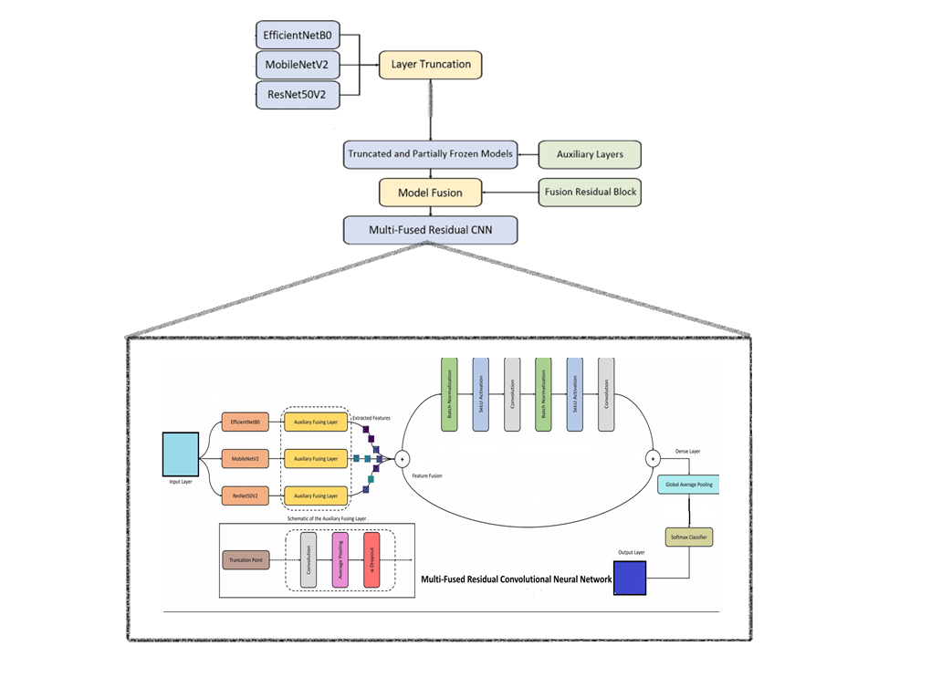
In this study, we proposed a novel Multi-Fused Residual Convolutional Neural Network (MFR-CNN) with Auxiliary Fusing Layers (AuxFL) to diagnose various types of brain tumor MRI images. The MFR-CNN was designed to handle four specific cases, namely glioma, meningioma, pituitary, and healthy brain images, obtained from reliable Kaggle databases. Our proposed model integrated three state-of-the-art models into a single feature extraction pipeline, incorporating partially frozen and truncated layers. This strategic fusion enabled the propagation of robust features and improved diagnostic performance without incurring significant computing costs, unlike most existing state-of-the-art models. Moreover, the MFR-CNN effectively mitigated overfitting and performance saturation issues, providing a notable advantage over models lacking these components. Upon evaluation, our proposed model achieved an outstanding accuracy of 94%, surpassing the efficiency and accuracy of conventionally trained DCNNs. Notably, the MFR-CNN demonstrated potential in enhancing brain tumor diagnosis more cost-efficiently than ensembles and outperforming conventional pre-trained and fine-tuned DCNNs. In conclusion, the proposed MFR-CNN with AuxFL and FuRB exhibits promising capabilities to improve the diagnosis of brain tumors, offering better cost-efficiency and accuracy compared to existing methods.
Downloads
Article Details

This work is licensed under a Creative Commons Attribution 4.0 International License.


